Courtesy of Eastwood et al, published in Advanced Energy Materials
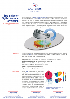
Digital Volume Correlation
Digital volume correlation (DVC) is a novel technique for full 3D strain and deformation measurements. The technique imports volume images of the component in reference and deformed states and is able to calculate the full 3D displacement and strain map. Images are typically acquired from X-ray Computed Tomography (X-ray CT) systems, but can equally be obtained by Magnetic Resonance Imaging (MRI) or Optical Coherence Tomography (OCT) systems for biological subjects, or via optical techniques for transparent media. DVC is a powerful nonintrusive technique for the identification of sub-surface material deformation and is capable of identifying defects, discontinuities or other material characteristics.
Digital volume correlation requires a random pattern within the bulk of the specimen. That random pattern is seen as changes in local contrast. In the case of X-ray CT scans the pattern will be produced by changes in material density such as air voids in concrete, or particles of different material type within the main body matrix, such as tin particles distributed in an aluminum powder The volume image is sub divided into interrogation sub-volumes within which the displacement of the pattern is computed.


Data courtesy of Dr. K. Madi / Prof. J. Tong (University of Portsmouth) et al., presented at ECCOMAS – Int. Conference on Tissue Engineering 2011, Lisbon